Pre-processing
Image pre-processing is essential to quantitative image analysis.
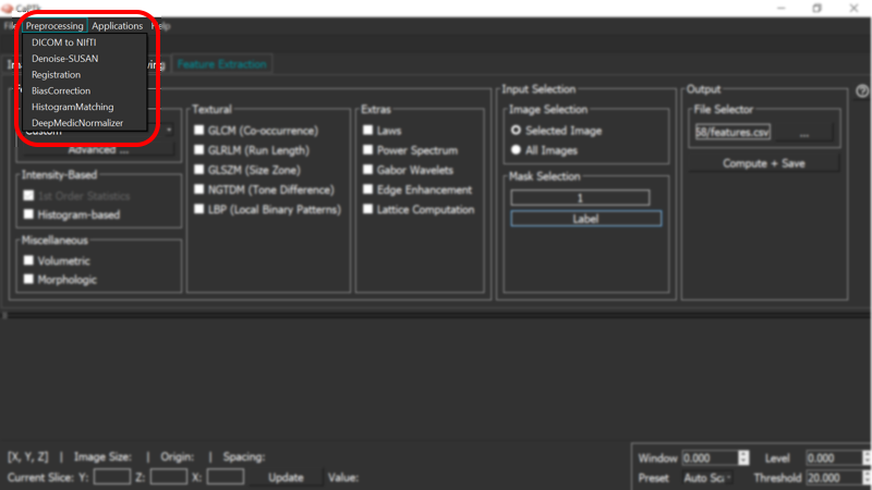
CaPTk pre-processing tools available under the "Preprocessing" menu are fully-parameterizable and comprise:
- Denoising. Intensity noise reduction in regions of uniform intensity profile is offered through a low-level image processing method, namely Smallest Univalue Segment Assimilating Nucleus (SUSAN) [1]. This is a custom implementation and does NOT call out to the original implementation distributed by FSL.
- Co-registration. Registration of various images to the same anatomical template, for examining anatomically aligned imaging signals in tandem and at the voxel level, is done using the Greedy Registration algorithm [5].
- Bias correction. Correction for magnetic field inhomogeneity is provided using a non-parametric non-uniform intensity normalization [2].
- Intensity normalization. Conversion of signals across modalities to comparable quantities using histogram matching [4].
- DeepMedic normalization. Images are normalized using a z-scoring mechanism with option to do the normalization within the region of interest or across the entire image. In addition, there is an option to remove outliers & noise from the image by removing a certain percentage of the top and bottom intensity ranges [6].
Available pre-processing algorithms
References:
- S.M.Smith, J.M.Brady, "SUSAN - a new approach to low level image processing", Int. J. Comput. Vis. 23(1):45-78, 1997. DOI:10.1023/A:1007963824710
- N.J.Tustison, B.B.Avants, P.A.Cook, Y.Zheng, A.Egan, P.A.Yushkevich, J.C.Gee, "N4ITK: Improved N3 Bias Correction", IEEE Trans Med Imaging. 29(6):1310-20, 2010. doi: 10.1109/TMI.2010.2046908
- S.Bauer, L.P.Nolte, M.Reyes, "Skull-stripping for Tumor-bearing Brain Images", arXiv. abs/1204.0357, 2012.
- L.G.Nyul, J.K.Udupa, X.Zhang, "New Variants of a Method of MRI Scale Standardization", IEEE Trans Med Imaging. 19(2):143-50, 2000. DOI:10.1109/42.836373
-
P.A.Yushkevich, J.Pluta, H.Wang, L.E.Wisse, S.Das, D.Wolk, "Fast Automatic Segmentation of Hippocampal Subfields and Medical Temporal Lobe Subregions in 3 Tesla and 7 Tesla MRI, Alzheimer's & Dementia: The Journal of Alzheimer's Association, 12(7), P126-127
- T.Rohlfing, N.M.Zahr, E.V.Sullivan, A.Pfefferbaum, "The SRI24 multichannel atlas of normal adult human brain structure", Human Brain Mapping, 31(5):798-819, 2010. DOI:10.1002/hbm.20906